
 Print This Post
Seeing as electrophysiology has taken up around 80% of my thoughts this past year, I thought it was only fitting to write a blog about it. In this blog, I am going to take you through what electrophysiology is, why it is such a powerful tool in neuroscience, and how I use it day-to-day during my PhD.
First off, what is electrophysiology and how does it work? Electrophysiology is a laboratory technique used to measure the electrical properties of cells and tissues. It allows us to detect how cells generate and respond to electrical signals by reading voltage changes in cell membrane and to measure current that flows through it. Electrophysiology approaches can help us to understand how excitable a cell is, how strongly it communicates with neighbouring cells, and how it’s activity changes in response to drugs, or in disease. In neuroscience, these recordings provide an insight into how neurons process information, both on their own and as a part of a network, and also how their function may become altered in disease.
As a part of my PhD, I do both whole-cell patch-clamp recordings, and extracellular field recordings. I use electrophysiology to study Alzheimer’s Disease, specifically how tau modulates ion channel dysfunction.
Whole-cell patch-clamp recordings allow me to study this at the level of an individual neuron. In this technique, a very fine glass microelectrode is bought into contact with the cell membrane and forms a high resistance seal. Once this seal is established, gentle suction is then applied to rupture the patch of membrane beneath the pipette tip, allowing us access to inside of the cell. In the lab, we call this “breaking through”. This configuration, known as the ‘whole cell mode’ allows us to directly measure the membrane voltage and ionic currents that flow across the neuronal membrane. It now becomes possible to measure how the neuron responds to electrical signals, how easily it fires action potentials, and how its membrane properties may change in health and disease.
I also use extracellular field recordings to look at this at a network level. I do these recordings in the hippocampus, the brain region responsible for learning and memory. In these recordings, I induce Long Term Potentiation, which is a form of synaptic plasticity involved in learning and memory – this is how we make long-term memories! To do this, I use one electrode to stimulate the Schaffer collateral pathway originating in the neurons in the CA3 region of the hippocampus, and a second electrode recording the response in the CA1 region. This allows me to assess how tau affects synaptic plasticity and how it may disrupt neuronal networks in a brain region critical for learning and memory.
For the purpose of this blog, I am going to focus primarily on whole-cell patch-clamp recording – the most technically difficult between these two techniques – and the first technique I learnt during my PhD.
I still remember the first time I successfully patched a cell. After weeks of broken pipettes and failed seals, I actually cried tears of joy when I saw that first stable recording appear on the screen. It sounds dramatic, but anyone who has learned patch clamp will probably understand exactly why.
Before any recording can begin, we have to run distilled water through the rig to make sure any residual salts have been removed from the tubing. Most of the time, the distilled water just flows in through the inflow tube, around the bath, out through the outflow tube, and into the waste. If there is a blockage – this is where the first hurdle of the day begins. Typically, you have to dismantle the tubing bit by bit until you find the source of the blockage, blast water through to unblock it, and then put everything back together again!
So now you have the inflow and outflow working, it’s time to turn the rig on. There are two types of recordings you can make, using voltage clamp and current clamp. In voltage clamp, you hold the cell at a particular voltage and record the current. In current clamp, you hold the cell at a particular current and record the voltage. Sounds simple, right? Well sometimes, the rig can ‘overload’, and it will be stuck in the wrong clamp. This can be for a number of reasons, usually because a tiny wire has gotten wet that’s not meant to! Once you have made sure: you’re in the right clamp, and you’ve got heated artificial cerebrospinal fluid (which we call aCSF) flowing through your rig, and nothings blocked – it’s time to get your tissue!
Something that every whole-cell patch-clamp electrophysiologist can relate to is learning how to find the pipette. The objective is submerged in aCSF, and you need to move the pipette into the liquid and focus on the tip of the pipette underneath the objective. I can safely say it took me and my friend and extremely long time, and countless broken pipette tips to successfully achieve this. Not that I’ve ever admitted this to anyone – but at the time I was learning, I actually had dreams about the pipette tip! Every new whole-cell patch-clamp electrophysiologist I meet, we always laugh and say, “do you remember how long it took to learn to find the pipette?” and compare times. In fact, my friend turned to me when we were learning, not joking, and said “Beccy, if I can never find the pipette, am I going to have to change PhD projects?”. We laugh about this now, and it’s on our ‘lab howlers’ list, and it seems like so long ago we were just starting out!
Everyone takes to whole-cell patch-clamp differently, and you have to have certain amount of resilience to master the technique. Me and my housemate learnt at the same time. At the time, it’s so hard not to compare yourself to others when you’re learning the same skill. It took me a little longer to get the hang of it than my housemate, and to be honest it was quite disheartening. Looking back on this now, you definitely shouldn’t compare yourself to others. We are all on our own individual journeys, and I am so glad I persevered – it was so worth it! Recently, teaching a new PhD student how to patch has reminded me exactly how steep that learning curve really is! Now, being able to sit in a row with my lab mates all patching side by side is one of my favourite parts of my PhD.
I’d say the bane of every electrophysiologist’s life is electrical noise. You can have your rig working perfectly one day, not move anything, and the next day you’re faced with so much electrical noise that you can’t record anything! You then put your hand on every single crocodile clip you can see to try and find the source of the noise. Sometimes, the noise just goes completely on its own – I think it is a phantom force!
My PhD supervisor always says that electrophysiology rigs are sent to test us – and I truly believe that!
Even when you finally spend what seems like the whole day troubleshooting the rig, and get everything working again, the next problem can be something as simple as discovering that the micropipette has been pulled just slightly too thick or thin and is the wrong shape to seal onto to the cell!
Something that I was really shocked over when I started electrophysiology was just how long you can record form a cell. Just a few days ago, my lab mate said she had been recording from a singular neuron for a whole hour. My supervisor always says, “every cell is a golden nugget”, and when a beautifully healthy neuron comes into view, says “that’s a beautiful one that is” – which has quickly caught on with all of us in the lab!
I’m lucky enough to have two amazing supervisors. They both make the lab environment such an enjoyable one to be in – that even if we’re having rubbish patching days and nothing is working as it should, we all still manage to have a laugh!
Like many lab techniques, it can be so easy sometimes to get carried away being sat on the rig in the ‘lab bubble’ and forget the bigger picture. In my opinion, being able to record individual neurons firing, and the electrical activity of a singular cell is one of the coolest things I have ever done. To think it will ultimately contribute to finding a treatment for Alzheimer’s disease is something that I feel so incredibly honoured to be able to do. I will leave you with a photo of me and the whole-cell patch-clamp rig!
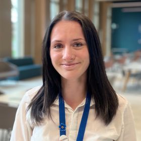
Beccy Owen
Author
Beccy Owen is a PhD Researcher at the University of Warwick, exploring how tau pathology disrupts neuronal ion channels and brain network activity in Alzheimer’s disease. As part of the Midlands Integrative Biosciences Training Programme, her work uses electrophysiology to better understand the molecular drivers of neurodegeneration. Originally from the Welsh countryside, Beccy’s passion for dementia research was shaped during her postgraduate studies and through personal experience with a family member living with the condition. She will be sharing her journey, insights, and lessons learned throughout her PhD here on the blog.


 Dementia Researcher
 Beccy Owen
 Bernie McInally
 Dr Chloe Fawns-Ritchie
 Dr Yvonne Couch
 Dr Clíona Farrell


 Rahul Sidhu

 Dr Emma Law
 Dr Ajantha Abey