
 Print This Post
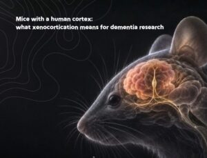 A Stanford-led team has transplanted human brain organoids into mice engineered to have almost no cortex of their own, producing the largest and best-integrated human neural graft reported so far. The unexpected bonus for anyone working on neurodegeneration: the grafts contained von Economo neurons, a cell type that has never appeared in a dish and that is lost early in frontotemporal dementia.
The study, from Konstantin Kaganovsky, Kevin Kelley, Tilo Gschwind and colleagues in Sergiu Pașca’s lab at Stanford, was published in Nature on 16 September and is open access. The authors call the technique xenocortication, and the logic behind it is simple enough to explain in a sentence: if you want human neurons to build something ambitious inside a mouse, first get the mouse’s own cortex out of the way.
Why human brain organoids needed more room
Previous transplantation work from the same group put human brain organoids into newborn rats, where they matured and wired into sensory pathways. But those grafts were squeezed in beside a rat cortex that was already growing fast, and rodent neurons develop far more quickly than human ones. A graft could reach about a third of a hemisphere, and it had to compete for every connection it made.
So the team built what they call an apallial mouse, deleting the cohesion factor Esco2 in cells expressing the cortical marker Emx1, on an immunodeficient background. The result is an animal that develops without most of its neocortex and hippocampus — MRI showed roughly half the normal brain tissue volume, and single-nucleus sequencing of 880,000 nuclei confirmed a sevenfold depletion of cortical excitatory neuron classes, with subcortical and olfactory populations preserved.
Four human brain organoids, grown from induced pluripotent stem cells, were then transplanted into each pup at around ten days old. Grafts took in 86% of 29 animals. Between two and three months post-transplant the human tissue grew 4.7-fold, and by three months it made up 91.9% of the total cortical volume, at roughly 32,000 neurons per cubic millimetre. That is a step change from a third of one hemisphere.
Wired in, and firing
The grafts were not passive lumps. Retrograde tracing found host inputs arriving from palaeocortex, thalamus and pallidum. Anterograde tracing followed human axons out of the graft, through subcortical structures, and — in every animal examined — as far as the cervical spinal cord, confirmed with a human-specific antibody. Grafts in mice with an intact cortex showed no such spinal projections.
Cortex-wide calcium imaging through optically cleared skulls showed large synchronous bursts propagating across the whole graft in around 100 milliseconds, lasting tens of seconds and recurring every few minutes, with electrode recordings confirming these were real network events rather than imaging artefacts. Graft activity tracked the animals’ orofacial movement. Transcriptionally, the human neurons sat at roughly late second trimester — this is developing cortex, not adult cortex.
Why this matters for neurodegeneration
The finding most likely to interest dementia researchers is a cell type. The grafts contained a distinct cluster of layer 5 extratelencephalic projection neurons carrying markers characteristic of human frontoinsular cortex, the region that houses von Economo neurons — large, bipolar or corkscrew-shaped cells found only in big-brained mammals and absent from rodents.
The authors then did something useful: they went looking for these cells in human brain organoids grown the conventional way, in a dish. Across 213,753 neurons in the Human Neural Organoid Cell Atlas, three scored above their threshold for this identity. Three. In the xenocortical grafts, 85% of the layer 5 cluster cleared it, and the grafts carried more than threefold the proportion seen in earlier transplantation approaches. Morphological analysis found cells with genuine von Economo shape in all three animals sampled, median soma diameter 29.8 micrometres against 9.3 for neurons generally.
That matters because von Economo neurons are selectively vulnerable. They are depleted in frontotemporal dementia, altered in the anterior cingulate across the lifespan and in Alzheimer’s disease, and implicated in schizophrenia and autism. Until now, anyone studying selective vulnerability in FTD had a choice between post-mortem human tissue and animals that do not possess the cell type at all. A living platform that generates them, in a body, with behaviour attached, is new ground.
What it cannot do yet
It is worth being clear about the limits, because the headline invites overreach.
The human brain organoids did not make the mice cleverer. Mice without a cortex failed a working memory task; the transplanted animals performed above chance, which is a partial rescue at best, and both groups were impaired on trace fear conditioning. Unsupervised analysis of spontaneous behaviour placed the transplanted animals between cortex-less and control mice but distinct from both, with markedly less stable behavioural repertoires. Gait was broadly normal with subtle differences in limb coordination.
The graft itself lacks canonical cortical layering, shows only weak areal patterning, and has very few inhibitory interneurons. The oldest organoids ran to 177 days. And the authors are candid that the mismatch between fast rodent development and slow human development may ultimately cap how far integration can go.
The disease application they actually demonstrated was perinatal hypoxia: five hours at 5% oxygen produced a HIF1α response confined to the human graft, vascular changes on susceptibility-weighted MRI ten days later, a microglial and astroglial reaction inside the graft, and measurable gait changes two days post-injury. That is a cerebral palsy model, not a dementia model. The route from here to modelling a late-onset neurodegenerative disease in human tissue inside a mouse is long, and would need grafts that are considerably older than these.
The ethics conversation, for once, arrives early
The work went through NIH guidelines and Stanford’s animal care panel, and the transplants were done days after birth, well after the mouse’s core wiring is established. The authors make the ethical case themselves rather than waiting to be asked: pushing towards grafts with mature layering, gyrification, balanced excitation and inhibition, or mature network properties — particularly if the developmental mismatch is closed by transplanting into embryonic hosts or non-human primates — will need guidance agreed in advance, with ethicists, patient representatives and regulators in the room.
That is the right instinct, and it is the part of this paper early career researchers should read closely. The technical achievement will be replicated and extended within a couple of years. The question of what a mostly-human cortex inside an animal means, and where the line sits, is one this generation of researchers will be asked to answer.
Kaganovsky, K., Kelley, K. W., Gschwind, T. et al. Developmental xenocortication using human-derived organoids in mice. Nature (2026). https://doi.org/10.1038/s41586-026-11032-2 — open access.




 Dr Ajantha Abey
 Dr Yvonne Couch