
One of the most satisfying moments as a researcher is when you look at your data and see that everything has worked exactly as you’d hoped it would. In my case, that was often indicated by taking a look down a microscope at a population of iPSC-derived neurons I had been working with for, often, several months. Weeks and weeks of carefully tending to them, changing media, following strict treatment regimes and meticulously sterilising everything (including myself) to ensure nothing gets contaminated. Finally witnessing the fruits of that labour is an incredibly satisfying thing.
When you work with neurons, the results can be especially beautiful. I did a lot of work with 3D cultures and really enjoyed sitting at a confocal microscope looking at long, branching processes that would stretch throughout the 3D matrix in all directions connecting like an intricate circuit. I particularly enjoyed calcium imaging which allowed me to see this 3D web of neural matter firing signals in a chain like an electrical circuit. It’s the sort of thing that would often remind me why I became a scientist in the first place.
But… there is an uncomfortable question that anyone working with neural cells should ask themselves regularly. If your cells ‘look’ like neurons, does that mean you have generated a good model of the brain? Not necessarily…
Over the years I have spent a lot of time in the lab working with stem cells. In my first postdoc, I spent four years working with neuronal differentiation to yield neurons that we could apply to making 3D models of neurodegenerative diseases. During that time, I learned that getting cells to look the way you want them to is only part of the challenge (more on that later).
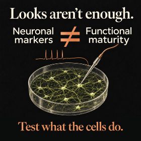
Neuronal shape and marker expression are useful evidence, but they do not establish functional maturity. A culture can produce a beautiful image without demonstrating the behaviour a study needs. As Sam Moxon argues, characterisation should also test what the cells actually do.
Before diving further into this, please allow me to load in a bit of background information for anyone who isn’t familiar with the type of work I am talking about. I was working with induced pluripotent stem cells (iPSCs). These are cells that have been extracted from skin biopsies and been reprogrammed into a state where they can develop into many different cell types. By exposing them to the right conditions, we can encourage them to become neurons and other cells found in the brain. They also carry the genetic ‘memory’ of the person they were extracted from. Therefore, we can take skin cells from a patient with Alzheimer’s disease, reprogramme them into iPSCs and then differentiate (or turn) them into brain cells that carry hallmarks of the issues that lie within the cells of that patient’s brain. This is potentially incredibly powerful because it can allow us to understand what’s happening inside human neurons without having to rely on post-mortem brain tissue.
Why iPSC-derived neurons need more than the right markers
However, producing something that resembles a neuron isn’t the same as producing something that behaves like one. You can get beautiful fluorescent images of iPSC-derived neurons with positive results for classic neuronal markers and they look lovely in a paper, but they only tell you so much. Specifically, they only show you that your cells have neuronal characteristics. It’s important but it is only part of the story. Are they mature? Are they the types of neurons we’re interested in studying? Do they possess the molecular machinery needed to perform their intended functions? And, perhaps most importantly, do they actually work?
There is one key thing that we have to pay a lot of attention to when working with iPSC-derived neural cells: functionality. We put a lot of effort into showing that our cells express the right markers. We use things like immunofluorescence microscopy, PCR or Western blots. It is all very sensible work but it’s missing something. A cell expressing neuronal markers doesn’t automatically mean it’s a functionally mature neuron.
A rainy day at Aston
This was something that really hit home to me on a rainy day at Aston University. I was working on my first postdoc at the University of Manchester and had come down to Aston to spend the day with Dr Eric Hill and his research team at Aston. Eric has always been a big advocate for ensuring the functionality of iPSC-derived neurons and I was very much in the trap of ‘just stain them and show they express the right stuff’. After chatting with Eric at a conference, he offered to test the functionality of the 3D neural networks I had been working on. A few weeks later I found myself sat in the lab, monitoring a hurricane off the coast of Florida hoping it wouldn’t affect our upcoming trip to Disney World (it didn’t). Suddenly I heard Dr James Crowe, one of Eric’s researchers, go ‘wow look at that’. I turned to see my neurons ‘talking to each other’. Firing a series of signals along their intricate network of processes and connections. I had never seen this before and it was mesmerising.
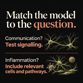
A useful model does not need to reproduce every feature of the human brain. It does need to represent the biological processes relevant to the question. A study of neuronal communication needs evidence of signalling; a study of inflammation needs the relevant cells and pathways. Choose and validate the model accordingly.
That moment fundamentally changed how I thought about my work. I was so focussed on making sure my cells looked like neurons that I hadn’t thought about whether they actually behaved like neurons. Now, there I was, watching activity in a network of cells I’d spent weeks carefully growing, and suddenly all those beautiful microscopy images seemed a little less impressive. Don’t get me wrong, they were still important, but this was an entirely different level of characterisation. And it’s still not enough…
I was so focussed on making sure my cells looked like neurons that I hadn’t thought about whether they actually behaved like neurons.
The images and videos were beautiful and they showed better evidence of creating a functional network but it still doesn’t come close to what happens inside our brains. Billions of neurons supported by an enormous variety of other cell types, all working together in an extraordinarily complex environment. There are astrocytes, microglia, blood vessels and countless interactions between different populations of cells. All of this is influenced by ageing, and all of it has a role to play in neurodegenerative disease. Our lab models cannot reproduce that complexity and that’s okay. Thanks to probably hundreds of thousands of hours of research the 3D models we can create in a dish are getting more and more complex and intricate. They still have significant limitations and are still a far cry from the human brain but they are, nonetheless, incredibly useful. We just need to be realistic about what they can tell us.
What makes iPSC-derived neurons a good model?
That’s the important point here. You don’t have to recreate every aspect of the human brain to create a good model but you do need to ensure you produce the ‘right’ biological processes. A model that looks pretty but isn’t functionally very useful carries less value than an ugly but functional one. If we’re investigating neuronal communication, we need evidence that our iPSC-derived neurons can communicate. If we’re studying the role of inflammation, we need to consider whether the relevant immune cells and signalling pathways are represented. The model needs to be appropriate for the question we’re asking, not simply impressive to look at. We need to build a picture using different types of evidence, from morphology and molecular markers to functional testing, and make sure our results are reproducible. Otherwise, we risk drawing conclusions about a disease from a model that isn’t behaving the way we think it is.
A model that looks pretty but isn’t functionally very useful carries less value than an ugly but functional one.
When I look back on that day at Aston, I still remember how exciting it was to see the activity of the iPSC-derived neurons in my samples. It didn’t make all the prior work meaningless but it did make me realise there was a lot more that I needed to consider. It’s a lesson that I have carried with me ever since. We should always be asking ourselves whether our models are actually capable of answering the questions we’re using them to investigate. After all, the goal isn’t to produce something that looks impressive on a conference slide. It’s to produce something we can trust.

Dr Sam Moxon
Author
Dr Sam Moxon is a Research Fellow at the University of Birmingham. His expertise falls on the interface between biology and engineering. His PhD focussed on regenerative medicine and he now works on trying to develop 3D bioprinting techniques with human stem cells, so that we better understand and treat degenerative diseases. Outside of the lab he hikes through the Lake District and is an expert on all things Disney.


 Adam Smith
 Dementia Researcher

 Beccy Owen

 Heather Wilkinson

 Dr Yvonne Couch






 Bernie McInally

 Dr Chloe Fawns-Ritchie